
ADVANCED COSMETIC PROCEDURES
ACP is an Advanced Cosmetic Procedure carried out by a qualified electrologist or a qualified medical practitioner.
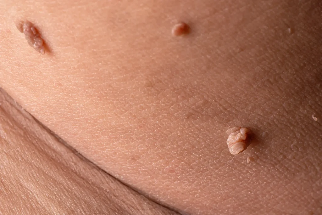
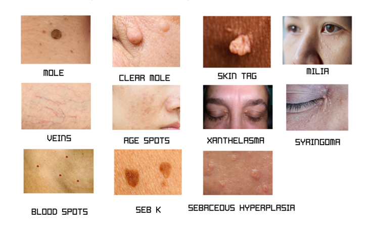
It can be used for the treatment of many superficial blemishes, including skin tags, warts, moles, thread veins, blood spots, milia, pigmentation (age spots/liver spots) and much more.
Advanced Electrolysis is a permanent and cost-effective treatment for the removal of these blemishes.
The NHS are no longer treating many of these blemishes, as they are classed as cosmetic treatments.
At Allure Beauty Clinic, we pride ourselves on staying at the forefront of all the latest advancements in all treatments, with continuous training to ensure all techniques are up to date, effective and safe.
Electrolysis has been used for over 140 years when in 1875, an ophthalmologist from Missouri, came up with a solution to remove painful ingrown eyelashes, and successfully used this for his patients. However, as with most inventions, other treatments are discovered which can give far more reaching benefits.
WHAT KIND OF BLEMISHES CAN BE TREATED?
Pigmentation/Sun spots/Age spots /Liver Spots
Pigmentation or age spots, also known as liver spots or sunspots, are flat, brown, gray, or black spots that appear on the skin. They are caused by an excess production of melanin, the pigment that gives skin its colour. These spots typically develop on areas of the skin that are frequently exposed to the sun, such as the face, hands, shoulders, and arms.
Causes of pigmentation and age spots
Sun Exposure: Overexposure to ultraviolet (UV) light from the sun is the primary cause of pigmentation/age spots. UV rays accelerate the production of melanin in certain areas of the skin, leading to the development of spots over time.
Aging: As we age, our skin’s ability to regenerate and repair itself decreases. This can result in the accumulation of pigmentation in certain areas, especially if there has been significant sun exposure over the years.
Hormonal Changes: Fluctuations in hormones, such as during pregnancy or due to certain medical conditions, can also contribute to the development of pigmentation spots.
HOW CAN WE TRY TO PREVENT PIGMENTATION/AGE SPOTS?
Sun Protection: The best form of prevention for pigmentation/age spots is to protect your skin from the sun. This includes:
- Using Sunscreen: Apply a broad-spectrum sunscreen with SPF 30 or higher every day, even on cloudy days.
- Wearing Protective Clothing: Cover exposed skin with hats, long sleeves, and sunglasses when outdoors.
- Avoiding Peak Sun Hours: Try to stay out of the sun during its peak hours, typically between 10 a.m. and 4 p.m.
Avoid Tanning Beds: Artificial UV radiation from tanning beds can also contribute to the development of pigmentation spots and skin damage. Avoid using tanning beds altogether.
Use of Topical Treatments: Certain topical treatments, such as those containing retinoids or hydroquinone, may help lighten existing pigmentation spots and prevent new ones from forming. However, these should be used under the guidance of a dermatologist.
Healthy Lifestyle: Maintaining a healthy lifestyle, including a balanced diet rich in antioxidants and vitamins, staying hydrated, and avoiding smoking, can also contribute to overall skin health and reduce the risk of pigmentation spots.
It’s important to note that while pigmentation spots are generally harmless, any new or changing spots should be evaluated by a dermatologist to rule out any underlying skin conditions or potential signs of skin cancer.
SPIDER NAEVI
Spider nevi, also known as spider angiomas or spider telangiectasias, are small, dilated blood vessels that appear close to the surface of the skin. They are named for their spider-like appearance, with a central red spot and radiating branches resembling spider legs. Understanding what spider nevi are, how they develop, and potential prevention strategies can help manage their occurrence.
Causes and Development of Spider Nevi:
Hormonal Changes: Spider nevi can be triggered by hormonal changes, such as those occurring during pregnancy, puberty, or hormonal imbalances.
Liver Dysfunction: Liver diseases, particularly those causing elevated estrogen levels (such as cirrhosis), can lead to the development of spider nevi.
Sun Exposure: While less common, excessive sun exposure and damage to the skin can contribute to the formation of spider nevi.
Genetic Predisposition: Some individuals may be more genetically prone to developing spider nevi.
Prevention of Spider Nevi:
Preventing spider nevi can be challenging, especially since some causes (like hormonal changes) are beyond individual control. However, certain strategies may help reduce their likelihood:
Protect Skin from Sun Exposure: Limiting sun exposure and using sunscreen (SPF 30 or higher) can help protect the skin and prevent damage that may contribute to the formation of spider nevi.
Healthy Lifestyle: Maintaining a healthy lifestyle, including regular exercise, a balanced diet, and avoiding excessive alcohol consumption, can support overall skin health and minimize factors like liver dysfunction that may contribute to spider nevi.
Seek Medical Evaluation: If spider nevi appear suddenly or are associated with other symptoms (such as liver disease), it’s important to seek medical evaluation and appropriate management by a healthcare professional.
Blood Spots/Cherry Angiomas
Cherry angiomas, also known as Campbell de Morgan spots or senile angiomas, are common skin growths made up of small blood vessels. They typically appear as smooth, cherry-red bumps on the skin’s surface. These benign lesions can vary in size, ranging from a pinhead to a quarter of an inch in diameter. While they are usually harmless and don’t require treatment, some individuals may choose to have them removed for cosmetic reasons or if they bleed or become irritated.
Causes and Development of Cherry Angiomas:
The exact cause of cherry angiomas is not fully understood, but they are believed to be linked to genetic factors and aging. The following factors may contribute to their development:
Aging: Cherry angiomas are more commonly seen in individuals over the age of 30, and their prevalence increases with advancing age.
Genetics: There may be a genetic predisposition to developing cherry angiomas, as they often run in families.
Hormonal Changes: Pregnancy and hormonal fluctuations can sometimes trigger the appearance of cherry angiomas.
Exposure to Chemicals: There is some evidence to suggest that exposure to certain chemicals or environmental factors may play a role in the development of cherry angiomas, although more research is needed in this area.
How and Where Cherry Angiomas Develop:
Cherry angiomas typically develop on the trunk of the body, including the chest, back, abdomen, and shoulders. However, they can also appear on other areas of the skin. These growths usually start as small red dots and may gradually increase in size over time. Some individuals may develop multiple cherry angiomas over the years.
Best Forms of Prevention and Management:
Cherry angiomas are generally harmless and do not require preventive measures. However, if you wish to minimize their appearance or prevent new ones from forming, consider the following tips:
Protect Your Skin: While there’s no direct way to prevent cherry angiomas, protecting your skin from excessive sun exposure and harmful chemicals can help maintain overall skin health.
Regular Skin Checks: Perform regular self-examinations of your skin to monitor any changes in existing cherry angiomas or the development of new ones. If you notice any unusual changes, consult a dermatologist for evaluation.
Cosmetic Removal: If cherry angiomas become bothersome due to bleeding, irritation, or cosmetic concerns, they can be removed by various methods, including laser therapy, ACP.
Seek Medical Advice: If you have concerns about cherry angiomas or notice any changes in your skin, consult a healthcare professional for proper evaluation and advice
Milia
Milia are small, benign cysts that appear as tiny white or yellowish bumps on the skin’s surface. They are often found on the face, particularly around the eyes, cheeks, and forehead, but can also occur on other parts of the body. Milia is commonly seen in newborns (referred to as “neonatal milia”) but can also affect individuals of any age, including adults.
Causes and Development of Milia:
The exact cause of milia can vary depending on the type. The two main types of milia are:
Primary Milia: These are formed when keratin (a protein found in skin tissues) becomes trapped beneath the skin’s surface, leading to the formation of small cysts. Primary milia can occur in individuals of any age.
Secondary Milia: These develop because of skin damage, such as burns, blistering, or prolonged sun exposure. Secondary milia can also occur after certain skin treatments, like laser resurfacing or dermabrasion.
How and When Milia Develop:
Milia develop when dead skin cells become trapped near the skin’s surface, forming small cysts. The following factors may contribute to their development:
Skin Care Products: Using heavy or oily skincare products that can clog pores.
Injury to the Skin: Skin damage from burns, sun exposure, or certain skin treatments.
Genetic Predisposition: Some individuals may be more prone to developing milia due to genetic factors.
Hormonal Changes: Hormonal fluctuations, particularly during pregnancy, can contribute to the formation of milia.
Best Forms of Prevention and Management:
Preventing and managing milia involves adopting certain skincare practices and, in some cases, seeking professional treatment. Here are some tips:
Gentle Skin Care: Use gentle cleansers and avoid heavy or greasy skincare products that can clog pores and contribute to milia formation.
Exfoliation: Regular exfoliation can help prevent the buildup of dead skin cells that can lead to milia. However, be gentle with exfoliation, as aggressive scrubbing can irritate the skin.
Sun Protection: Protect your skin from excessive sun exposure by wearing sunscreen daily and avoiding prolonged sun exposure, which can contribute to skin damage and secondary milia.
Professional Treatment: In clinic by using ACP methods.
Seborrheic Keratosis (SK)
Seborrheic keratosis (SK) is a common non-cancerous skin growth that appears as a waxy, scaly, or raised lesion. These growths can vary in color from light tan to dark brown or black and may have a slightly elevated or rough surface. Seborrheic keratoses are typically found on areas of the skin exposed to the sun, such as the face, chest, shoulders, back, or other sun-exposed areas.
Causes and Development of Seborrheic Keratosis:
The exact cause of seborrheic keratosis is not known, but several factors may contribute to its development:
Aging: Seborrheic keratoses are more common with advancing age, typically appearing after the age of 40. They may increase in number and size as individuals get older.
Sun Exposure: UV radiation from sunlight may play a role in the development of seborrheic keratoses, as they often appear on sun-exposed skin.
Genetics: There may be a genetic predisposition to developing seborrheic keratoses, as they can run in families.
How and When Seborrheic Keratosis Develops:
Seborrheic keratoses develop gradually over time and can vary in appearance. They may start as small, flat spots and gradually thicken, develop a waxy texture, and become raised. These growths are typically painless and may appear singly or in clusters.
Best Forms of Prevention and Management:
Preventing seborrheic keratosis is not always possible, but you can take steps to manage and monitor them:
Sun Protection: Protect your skin from excessive sun exposure by wearing sunscreen daily with SPF 30 or higher, seeking shade, and wearing protective clothing.
Regular Skin Checks: Perform regular self-examinations of your skin to monitor for any new or changing skin growths, including seborrheic keratoses.
Treatment Options for Seborrheic Keratosis:
We use ACP in the clinic and have amazing results.
We will always advise having blemishes checked by a GP prior to any treatment.
MOLES
Moles, also known as nevi, are common skin growths that appear as small, dark spots or patches on the skin. They are usually brown in color but can also be tan, black, red, pink, or flesh-colored. Moles can vary in size, shape, and texture, and most are benign (non-cancerous). Moles are clusters of pigmented cells called melanocytes, which give them their characteristic color.
Causes and Development of Moles:
The development of moles is primarily influenced by genetic factors and sun exposure. The following factors may contribute to mole development:
Genetics: Some individuals are more predisposed to developing moles due to inherited genes.
Sun Exposure: Sun exposure can trigger the development of moles, especially in fair-skinned individuals.
Hormonal Changes: Hormonal changes during puberty, pregnancy, or with hormonal therapies can stimulate the appearance of new moles.
How and When Moles Develop:
Moles can develop at any age, but are mostly acquired during childhood and adolescence. They can continue to appear throughout adulthood, with newmoles typically appearing until the age of 40. Moles can develop on any part of the body, including areas not exposed to the sun.
Best Forms of Prevention and Management:
While it’s not always possible to prevent the development of moles, you can take steps to monitor and manage them effectively:
Sun Protection: Protect your skin from excessive sun exposure by wearing sunscreen with SPF 30 or higher, seeking shade, and wearing protective clothing, especially if you have many moles.
Regular Skin Checks: Perform regular self-examinations of your skin to monitor for any changes in moles, such as changes in size, shape, color, or texture.
Avoidance of Tanning Beds: Avoid using tanning beds and sunlamps, as they can increase the risk of developing moles and skin cancer.
Monitor Moles for any Change.
It’s important to keep an eye on your moles and seek medical evaluation if you notice any of the following changes, which could indicate skin cancer (melanoma):
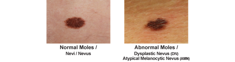
Changes in size, shape, or color of the mole
Irregular or uneven borders
Itching, bleeding, or crusting
Multiple colors within the mole
Evolution or change in appearance over time
No treatment will be performed without written consent from a GP.
FAQ’S
1. HOW DOES IT WORK?
Advanced Electrolysis uses a Thermolysis current and a very fine needle, which either cauterizes or lifts away the blemish.
2. HOW MANY TREATEMENTS WILL I NEED?
Usually 1 treatment is needed, with a follow up appointment to review.
3. HOW LONG DOES THE PROCEDURE TAKE?
The procedure itself can take seconds to a few minutes for 1 blemish, but this will be discussed at your consultation.
4. DOES IT HURT?
Everyone has a different pain threshold; however most people explain it to feel like pin pricks. If you feel like you would like to use a topical numbing cream, you can do this 30 min prior to your appointment.
5. WHAT HAPPENS AFTER THE TREATMENT?
You will be advised of aftercare verbally and given an aftercare sheet to follow, to ensure your skin repairs itself effectively. Healing can take between a few days and 2 weeks.
6. HOW WILL THE SKIN LOOK AFTER THE TREATMENT?
Your skin may be red and swollen immediately after. There may be some itching especially as the skin heals. But this is perfectly normal.
7. I AM TAKING MEDICATION; WILL I BE SUITABLE FOR TREATMENT?
Medications used for blood disorders including, blood thinners will not be suited to this treatment.
Those who have a pacemaker are also not suitable.
Also, if you suffer with keloid scarring this treatment would not be suitable.